Involvement of PRIP in mouse reproduction system
(Lab Mol Cell Biochem, Grad School of Dent Sci, Kyushu University)
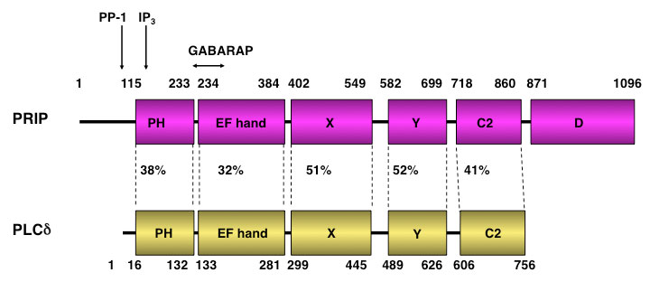
*A novel inositol 1,4,5-trisphosphate binding protein was identified.
The molecule is similar to phospholipase C (PLC)-_1 which has
catalytic X and Y domains and several domains such as PH, EF hand and C2.
However it lacks the catalytic activity.
Therefore we named PRIP (PLC-related catalytically inactive protein).
PRIP has two isoforms, PRIP-1 and PRIP-2.
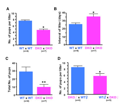
Figure 1. The fertility of WT and PRIP deficient (DKO) mice.
Wild type (WT) and PRIP-1, -2 double knockout (DKO) females (12 week-old) were bred with age-matched WT or DKO males for 4 months. The number of pups per litter, litters per female and interval of litter were scored (Student’s t test; *P < 0.05, **P < 0.01).

Figure 2. Body and tissues weights of WT and DKO mice.
Ovary, testis, uterus and pituitary gland were isolated after determining body weight, and were weighed. *P < 0.05.
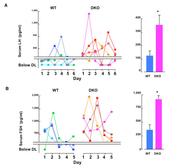
Figure 3. Serum LH and FSH levels.
Blood samples were obtained from 3 month-old female WT and DKO mice at 1700 h each day, and the serum were prepared. Serum LH and FSH levels were measured by ELISA. Each color on graph indicates the value of individual mice. The histograms shown on the right represent the average of serum gonadotropin levels measured in this experiment (Student’s t test; *P < 0.05).
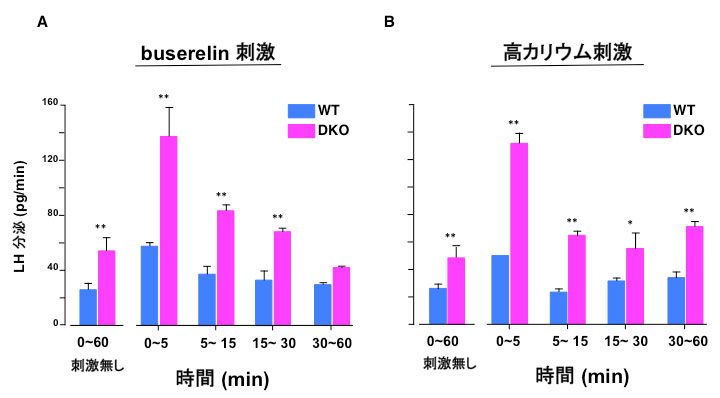
Figure 4. Serum LH and FSH levels.
Blood samples were obtained from 3 month-old female WT and DKO mice at 1700 h each day, and the serum were prepared. Serum LH and FSH levels were measured by ELISA. Each color on graph indicates the value of individual mice. The histograms shown on the right represent the average of serum gonadotropin levels measured in this experiment (Student’s t test; *P < 0.05).
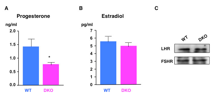
Figure 7. Serum levels of sex steroid hormones and the expression of gonadotropin receptors.
Same serum samples of WT and DkO as shown in Fig. 4 were used for the measurement of progesterone (A) and estradiol (B) by ELISA. The average of the measurements were shown as graphs. *P < 0.05. WT and DKO ovary extracts were analyzed for gonadotropin receptors by immunoblotting using indicated primary antibodies (C).
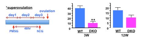
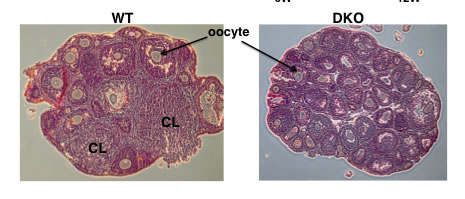
Figure 8. Numbers of ovulated oocytes according to female genotype
Pregnant-mare serum gonadotropin (PMSG) was injected to 3 and 12 week-old female mice at 1800 h on day 1 and chronic gonadotropin (hCG) was injected on day 3. Ovulated oocytes were collected from the oviduct and quantified on day 4. Histological data shows ovaries after ovulation. Paraffin-embedded sections of WT and DKO ovaries were stained with hematoxylin-eosin. CL: corpus luteum, n=6 respectively.
The characterization of PRIP-DKO mice is:
・fewer pups per litter and some of mutant pairs delivered no pups
・prolonged interval between litter occasions
・the reproductive defect was rather in females
・female mice exhibited a smaller uterus
・estrous days were increased
・serum levels of LH and FSH were increased
・pituitary culture exhibited the elevated basal level of LH secretion
・anterior lobe secreted more LH in response to buserelin and high K+
・serum progesterone level were apparently lower
・the amount of LH and FSH remained in pituitary were decreased
・the number of the ovulated oocytes much decreased
Home