|
|
|
|

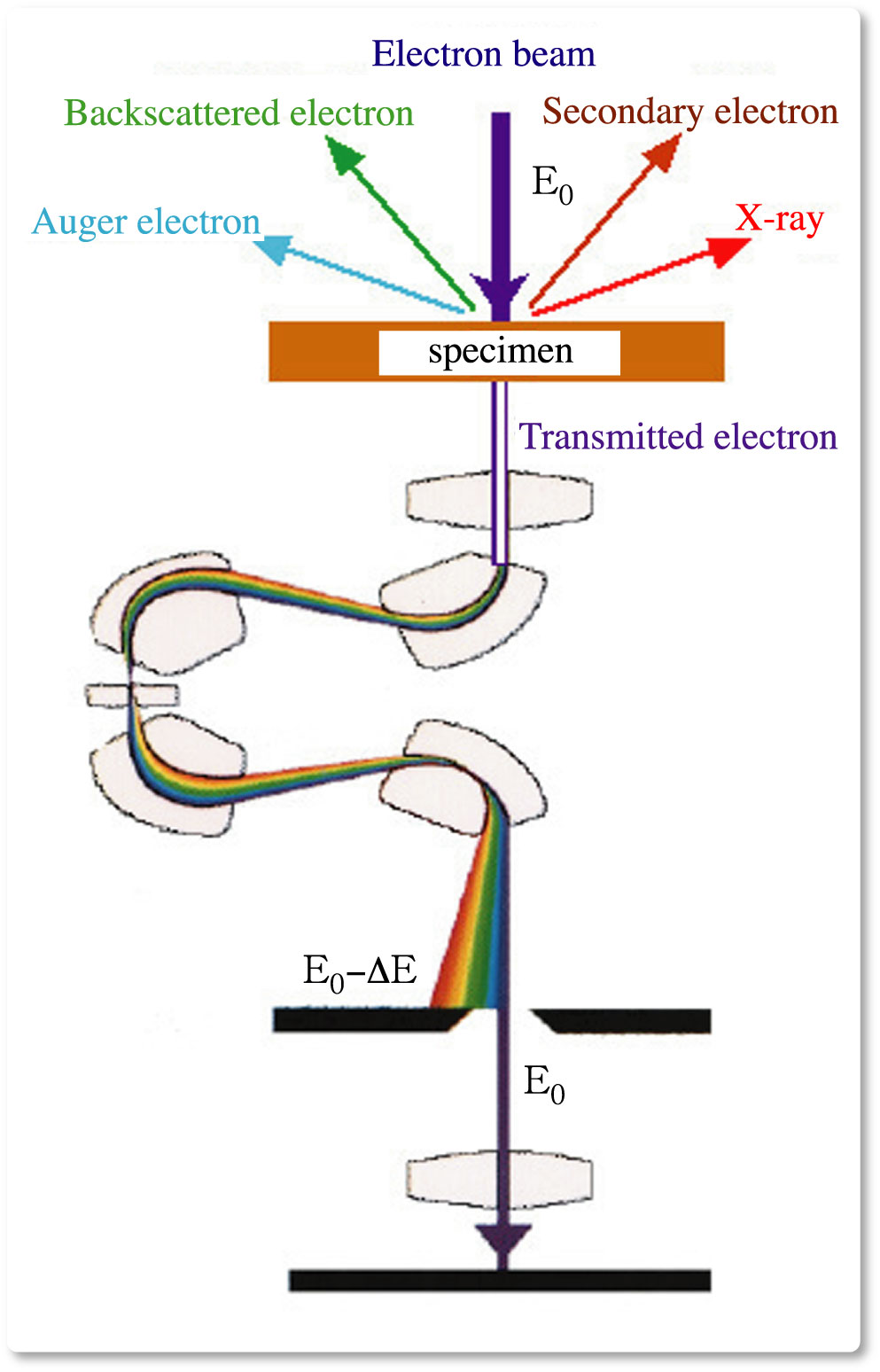
When a high-energy electron beam traverses a sample, many interactions occur between the beam electrons and the electrons in the material; for imaging purposes one is usually interested in the elastic part of the scattered electrons, and necessary to remove the inelastically scattered electrons from the images. Inelastic scattering electrons, i.e., the incident electron looses a fraction of its energy, result in blurry images and a decreased signal-to-noise ratio. To overcome such dilemma, numbers of researchers have worked on the development of energy-filters since the early stages of transmission electron microscope. In energy-filtered TEM, the electron microscopist selects electrons that have lost a certain amount of energy in inelastic scattering processes, and creates an image with those electrons. Since the energy-loss spectrum of a material contains a signature of all the chemical species present, one can actually "tune in" to a certain element and obtain the two-dimensional distribution of elements in the specimen and select optimum image contrast. Energy shifts can be observed which are related to the chemical environment of the atoms, and hence bonding information may be derived from the fine structure of the energy-loss spectrum.
|